What is Trochanteric Bursitis?
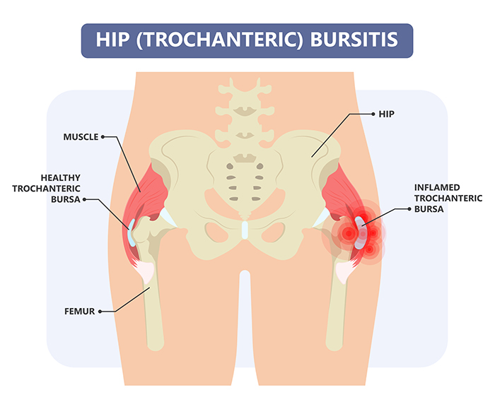
The hip has two large bursae. Bursae are jelly-like sacs located throughout the body that are positioned between bones and soft tissue to reduce friction.
One bursa covers the bony point of the hip bone called the greater trochanter. Inflammation of this bursa is called trochanteric bursitis.
The iliopsoas bursa is found on the groin side of the hip between the anterior joint capsule and the iliopsoas tendon
Causes of Trochanteric Bursitis
Trochanteric bursitis may result from one or more of the following events:
- Injury to the point of the hip – including falling onto the hip, bumping the hip into an object, or lying on one side of the body for an extended period
- Overuse due to recreational, sporting, or work activities causing friction to the bursa on the side of the hip – including running up stairs, climbing, or standing for long periods of time
- Incorrect posture – resulting from scoliosis, arthritis of the lumbar spine, and other spinal problems
- Soft Tissue Stress – abnormal or poorly positioned joint or bone (leg length discrepancies or arthritis in a joint)
- Other Conditions – like rheumatoid arthritis, gout, and psoriasis,
- Previous Hip Surgery– may cause scar tissue that can contribute to bursitis
- Bone Spurs – or calcium deposits in the tendons that attach to the trochanter.
- Abductor tendon degeneration at their point of attachment into the greater trochanter
Bursitis is more common in women and in middle-aged or elderly people. In many cases, however, the cause of trochanteric bursitis is unknown.
Symptoms of Trochanteric Bursitis
Typically, trochanteric bursitis causes pain around the greater trochanter at the side of the hip:
- At the point of the hip, outside of the hip and thigh, and in the buttock
- When walking upstairs or squatting
- During activities such as getting up from a deep chair
- When lying on the affected side
- When pressure is on the outside of the hip
In the early stages the pain is usually described as sharp and intense. Later on the pain may become more of an ache and spread across a larger area of the hip and lateral thigh.
Diagnosis of Trochanteric Bursitis
The diagnosis is usually made by:
- Taking a medical history
- Performing a physical examination
- Imaging tests
Imaging Tests
- X-rays may show calcification and bony spurs at the trochanter
- MRI scans will usually show fluid within the inflamed bursa and are also useful to show any degeneration of the abductor muscles and tendons that attach to the trochanter
- Ultrasound scans may show fluid in the bursa
Treatment of Trochanteric Bursitis
Treatment goals include reducing pain and inflammation, preserving mobility, and preventing disability and recurrence. Treatment recommendations may include a combination of
- Rest
- Non-steroidal anti-inflammatory drugs.
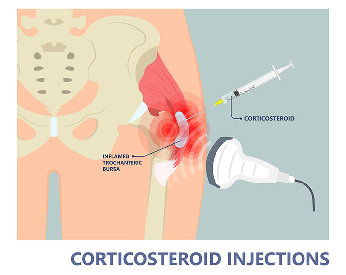
- Corticosteroid injections- can work quickly to decrease the inflammation and pain
- Physical therapy that includes a range of motion exercises and stretching of the iliotibial band and stretching of muscles around the hip is the mainstay for the treatment.
- Surgery is rarely required but may be recommended when other treatments are not effective or when there is tearing and degeneration of the gluteal muscle tendons that attach to the greater trochanter.
Trochanteric Bursitis Surgery
Recalcitrant trochanteric bursitis can be treated with bursectomy and iliotibial band (ITB) release.
Occasionally, a formal abductor repair may be required if a tear in the gluteal tendons is present.
Removal of the trochanteric bursa does not damage the hip, and the hip can function normally without it.
This is performed via a minimally invasive approach by removing the bursa through a small incision over the hip.