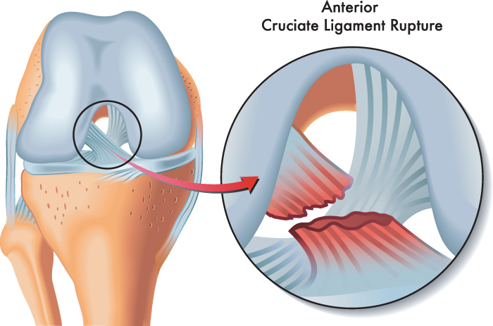
Anterior Cruciate Ligament (ACL)
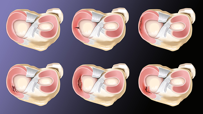
Anterior Cruciate Ligament (ACL) Tear The anterior cruciate ligament is a thick rope like structure made from collagen that joins the femur to the tibia. Its main function is to provide rotational stability to a knee joint. The anterior cruciate ligament is one of the major stabilising ligaments in the knee. It acts as a …